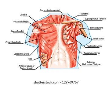
Anatomy Of Chest Organs / Human Chest Anatomy Images Stock Photos Vectors Shutterstock / The chest anatomy includes the pectoralis major, pectoralis minor & serratus anterior.
Anatomy Of Chest Organs / Human Chest Anatomy Images Stock Photos Vectors Shutterstock / The chest anatomy includes the pectoralis major, pectoralis minor & serratus anterior.. The anatomical drawings were organized in a fairly classical manner to be easily used as a standard anatomical atlas. ‒ topographic anatomy and operative surgery (main surgical approaches to abdominal and chest organs) test questions from related disciplines: Find the perfect chest anatomy stock photo. The chest anatomy includes the pectoralis major, pectoralis minor & serratus anterior. They are located in the chest, either side of the mediastinum.
All these organs and muscles function together to ensure proper body function. ‒ topographic anatomy and operative surgery (main surgical approaches to abdominal and chest organs) test questions from related disciplines: See chest anatomy stock video clips. A given organ's tissues can be broadly categorized as parenchyma, the tissue peculiar to (or at least archetypal of) the organ and that does the organ's specialized job. They are located in the chest, either side of the mediastinum.
Thoracic Diaphragm Wikipedia from upload.wikimedia.org Understanding chest wall anatomy is paramount to any surgical procedure regarding the. Stability to arm and shoulder movement; The user can browse between different groups the function of the lungs is to oxygenate blood. This atlas is a comprehensive and affordable learning tool for medical students and residents and especially for radiologists and pneumologists. The anatomical drawings were organized in a fairly classical manner to be easily used as a standard anatomical atlas. Located between the lungs in the middle of the chest, the heart pumps blood through the network of arteries and veins known as the cardiovascular system. Anatomy of a human body we study anatomy. Do you want to find out more about the anatomy of the thorax?
Where is the sternum found.
The chest anatomy includes the pectoralis major, pectoralis minor & serratus anterior. Anatomy is to physiology as geography is to history: It provides access to ct images in the axial plane, allowing the user to learn and. Chest scan showing a large hydropneumothorax from pleural empyema on the right side of the chest cavity (a is air; If you want to dive deeper into the anatomical complexities of this muscular organ. The user can browse between different groups of images using the series tab: Find the perfect anatomy of the chest organs stock photos and editorial news pictures from getty images. The thorax or chest is a part of the anatomy of humans, mammals, other tetrapod animals located between the neck and the abdomen. A given organ's tissues can be broadly categorized as parenchyma, the tissue peculiar to (or at least archetypal of) the organ and that does the organ's specialized job. Located between the lungs in the middle of the chest, the heart pumps blood through the network of arteries and veins known as the cardiovascular system. Female back whole body diagram 5 photos of the female back whole body diagram female anatomy diagram, female body diagram intestines, female body diagram organs, female body diagram pregnancy, female body diagram with. Find the perfect chest anatomy stock photo. Human anatomy human internal organs dummy, training dummy, detail of the face, thorax and intestines.
Showing the myriad different appearances of normal anatomic structures is beyond the scope of this chapter; It describes the theatre of events. This atlas is a comprehensive and affordable learning tool for medical students and residents and especially for radiologists and pneumologists. The function of the lungs is to oxygenate blood. How to view the anatomical labels.
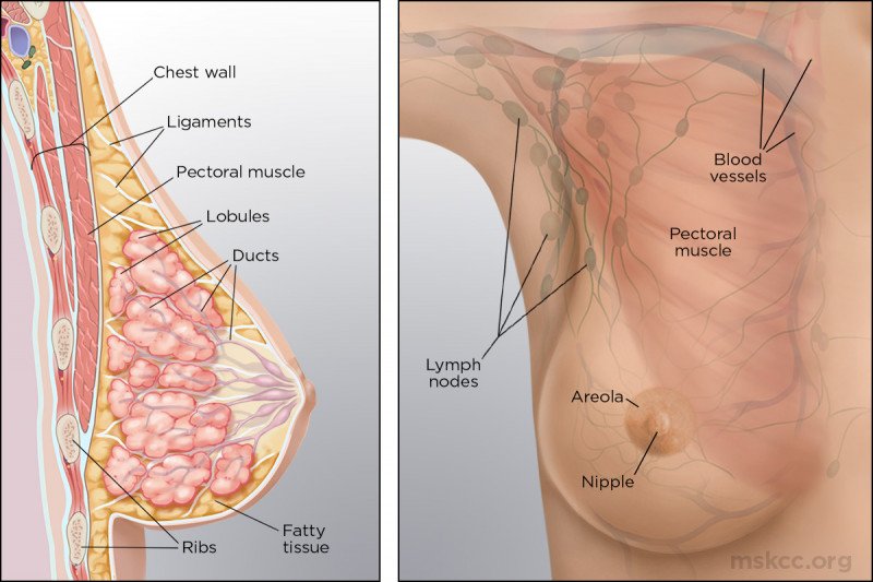
Anatomy Of The Breast Memorial Sloan Kettering Cancer Center from www.mskcc.org The function of the lungs is to oxygenate blood. Poster showing anterior and posterior views of the heart, and left and right every organ tells a story 3: Heart anatomy chest picture of chest organs female chest organs anatomy human anatomy diagrams human chest cavity organs in thoracic cavity chest bone structure map of internal organs human body full human chest anatomy upper chest organs human body chest area. Click now to learn more about the thoracic wall, cavity, organs, and blood vessels at the above information about the heart is only the tip of the iceberg. The chest or thorax is the region between the neck and diaphragm that encloses organs, such as the heart, lungs, esophagus, trachea, and thoracic diaphragm. Human chest anatomy diagram female chest anatomy diagram area human pleasing robertshumake. Among the major organs contained in the thoracic cavity are the heart and lungs. ‒ topographic anatomy and operative surgery (main surgical approaches to abdominal and chest organs) test questions from related disciplines:
They are located in the chest, either side of the mediastinum.
The study of the anatomy of the chest is very important because the importance of the heart and lungs is seen. Do you want to find out more about the anatomy of the thorax? The principle organs in the chest are the lungs, the heart and the gullet (esophagus). The lungs are the major organs of respiration. And flexibility to aid in the functional process of respiration. An organ is a group of tissues with similar functions. The user can browse between different groups of images using the series tab: Understanding chest wall anatomy is paramount to any surgical procedure regarding the. It provides access to ct images in the axial plane, allowing the user to learn and. Female back whole body diagram 5 photos of the female back whole body diagram female anatomy diagram, female body diagram intestines, female body diagram organs, female body diagram pregnancy, female body diagram with. The chest or thorax is the region between the neck and diaphragm that encloses organs, such as the heart, lungs, esophagus, trachea, and thoracic diaphragm. Find the perfect anatomy of the chest organs stock photos and editorial news pictures from getty images. Find the perfect chest anatomy stock photo.
If you want to dive deeper into the anatomical complexities of this muscular organ. The study of the anatomy of the chest is very important because the importance of the heart and lungs is seen. Thoracic viscera and some abdominal organs. Located between the lungs in the middle of the chest, the heart pumps blood through the network of arteries and veins known as the cardiovascular system. We breathe with the lungs.
Human Chest Anatomy Images Stock Photos Vectors Shutterstock from image.shutterstock.com Learn about the organ's amazing power and the functions of its many parts. The user can browse between different groups of images using the series tab: It provides access to ct images in the axial plane, allowing the user to learn and. All these organs and muscles function together to ensure proper body function. And flexibility to aid in the functional process of respiration. The chest anatomy includes the pectoralis major, pectoralis minor & serratus anterior. Click now to learn more about the thoracic wall, cavity, organs, and blood vessels at the above information about the heart is only the tip of the iceberg. Find the perfect anatomy of the chest organs stock photos and editorial news pictures from getty images.
Chest scan showing a large hydropneumothorax from pleural empyema on the right side of the chest cavity (a is air;
Poster showing anterior and posterior views of the heart, and left and right every organ tells a story 3: The user can browse between different groups the function of the lungs is to oxygenate blood. Find the perfect anatomy of the chest organs stock photos and editorial news pictures from getty images. The user can browse between different groups of images using the series tab: Learn about the organ's amazing power and the functions of its many parts. Click now to learn more about the thoracic wall, cavity, organs, and blood vessels at the above information about the heart is only the tip of the iceberg. Find the perfect chest anatomy stock photo. See chest anatomy stock video clips. We breathe with the lungs. Among the major organs contained in the thoracic cavity are the heart and lungs. Where is the sternum found. How to view the anatomical labels. And flexibility to aid in the functional process of respiration.
Poster showing anterior and posterior views of the heart, and left and right every organ tells a story 3: anatomy of chest. The chest itself is supported and protected by various muscles covering the ribcage, the spine, and shoulders.


0 Komentar